Types of Dental Radiographs and their Uses
Types of Dental Radiographs and their Uses
Dental radiography, also known as dental X-ray imaging or radiology, is a diagnostic technique used by dentists to visualize and assess various aspects of a patient’s oral health. It involves the use of X-ray technology to create images of the teeth, jawbone, and surrounding structures. Dental radiographs are essential tools that help dentists diagnose dental problems, plan treatments, and monitor oral health over time.
Types of Dental Radiographs
Here are the most common types and their uses:
Bitewing X-rays:
Use: Bitewing X-rays are primarily used to detect dental caries (cavities) between teeth and to assess the height of the alveolar bone, which supports the teeth.
Appearance: These X-rays show the crowns of the upper and lower teeth, with a clear view of the areas between them.
Periapical X-rays:
Use: Periapical X-rays provide a detailed view of a specific tooth from the crown to the root tip. They are used to identify issues such as tooth fractures, infections, abscesses, and bone loss around the tooth.
Appearance: Periapical X-rays capture the entire tooth from the crown to the root, as well as the surrounding bone.
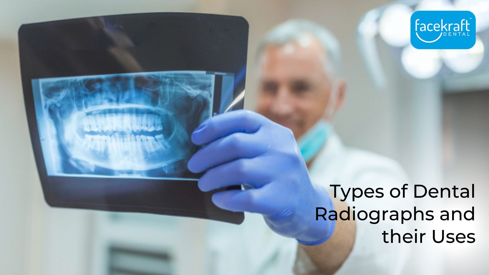
Panoramic X-rays:
Use: Panoramic X-rays provide a broad view of the entire mouth, including the teeth, jaws, and temporomandibular joints. They are used to assess impacted teeth, jaw disorders, and general oral health.
Appearance: These X-rays create a single, panoramic image of the entire oral cavity.
Occlusal X-rays:
Use: Occlusal X-rays are primarily used to visualize large areas of the upper or lower jaw. They are often used in paediatric dentistry to assess the development of primary and permanent teeth.
Appearance: Occlusal X-rays capture a top-down view of the upper or lower jaw, showing the arrangement of teeth and their relation to one another.
Cephalometric X-rays:
Use: Cephalometric X-rays are used by orthodontists to assess the overall facial and dental structures. They help in orthodontic treatment planning and evaluating facial growth.
Appearance: Cephalometric X-rays capture a side view of the head, showing the entire skull, jaw, and teeth.
Cone Beam Computed Tomography (CBCT):
Use: CBCT provides detailed 3D images of the teeth, jawbone, and surrounding structures. It is used for complex dental procedures like implant placement, orthodontic treatment planning, and assessing pathology.
Appearance: CBCT images are three-dimensional and offer a cross-sectional view of the oral and maxillofacial region.
Intraoral X-rays:
Use: Intraoral X-rays encompass a range of X-rays taken inside the mouth. They include bitewing, periapical, and occlusal X-rays, providing detailed views of teeth and surrounding structures.
Appearance: Intraoral X-rays capture close-up images of individual teeth or small sections of the mouth.
Extraoral X-rays:
Use: Extraoral X-rays are taken from outside the mouth and include panoramic and cephalometric X-rays. They are used to assess larger areas of the oral and maxillofacial region.
Appearance: Extraoral X-rays provide broader, overall views of the oral and facial structures.
Dental radiographs play a crucial role in diagnosing and monitoring oral health conditions. The best dentists use these images to detect problems early, plan treatments, and track the progress of dental procedures. The choice of radiograph type depends on the specific diagnostic needs of the patient.